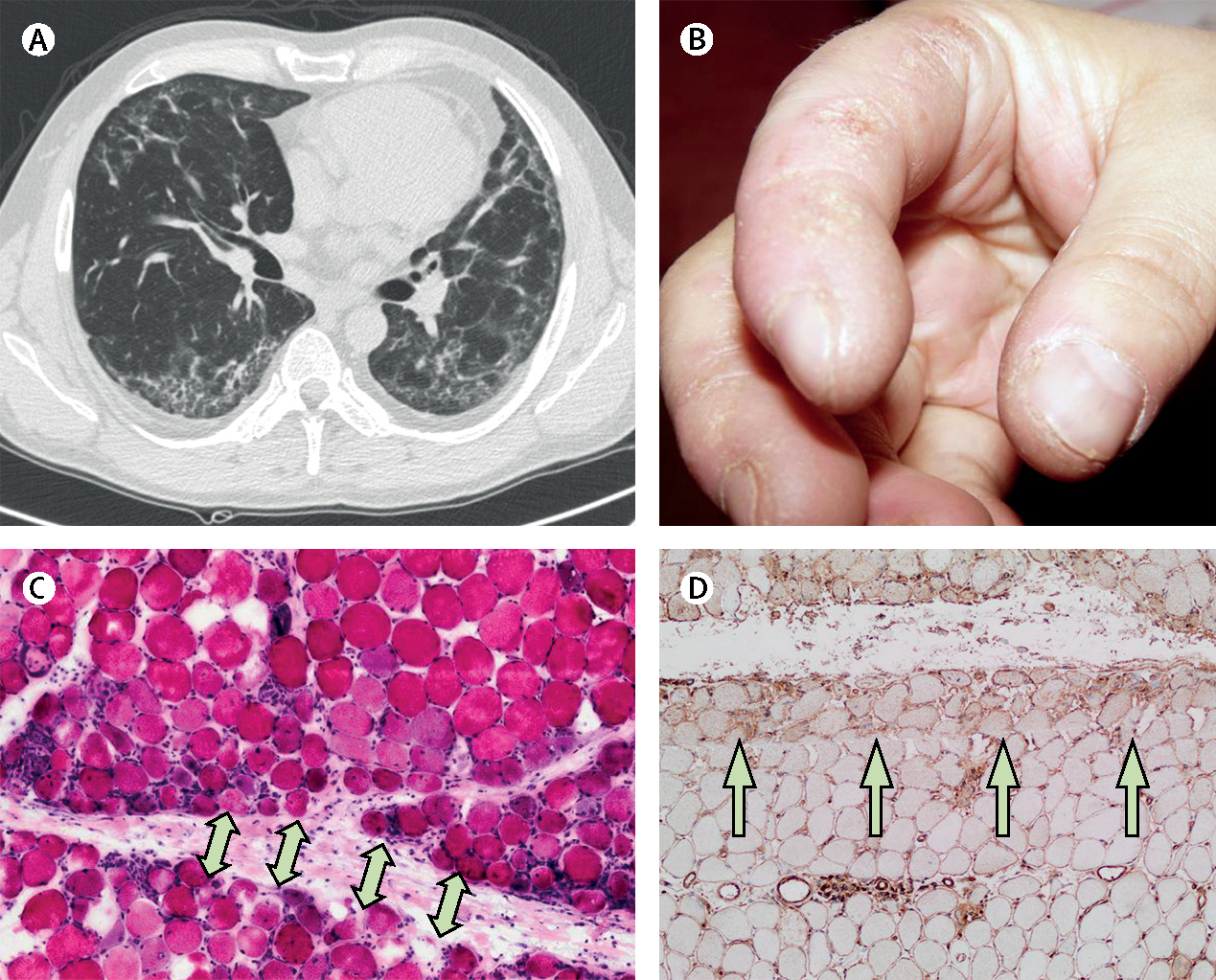
Figure 4: Clinical features and radiological and pathological findings of antisynthetase syndrome.

A woman aged 45 years presented with muscle weakness and dyspnoea. (A) A high-resolution chest CT scan showed interstitial lung disease. She had crackles in both lung bases and (B) mechanic’s hands. Muscle biopsy showed (C) necrotic and regenerating muscle fibres in the perifascicular area (arrows) and (D) prominent class-1 major histocompatibility complex positivity predominantly in the perifascicular area (arrows). Serum was positive for anti-Jo1 antibodies. Jo1=histidyl tRNA synthetase.
