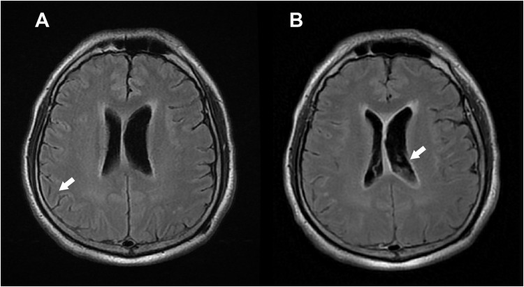
Figure 1.
Magnetic resonance imaging of the brain (A) Leptomeningeal enhancement (white arrow) in bilateral cerebral sulci and brainstem surface. (T2-weighted Fluid-attenuated inversion recovery [T2-FLAIR]) (B) Subependymal enhancement with sedimentations (white arrow) in bilateral lateral ventricles. (Contrast-enhanced Fluid- attenuated inversion recovery [CE-FLAIR]).

