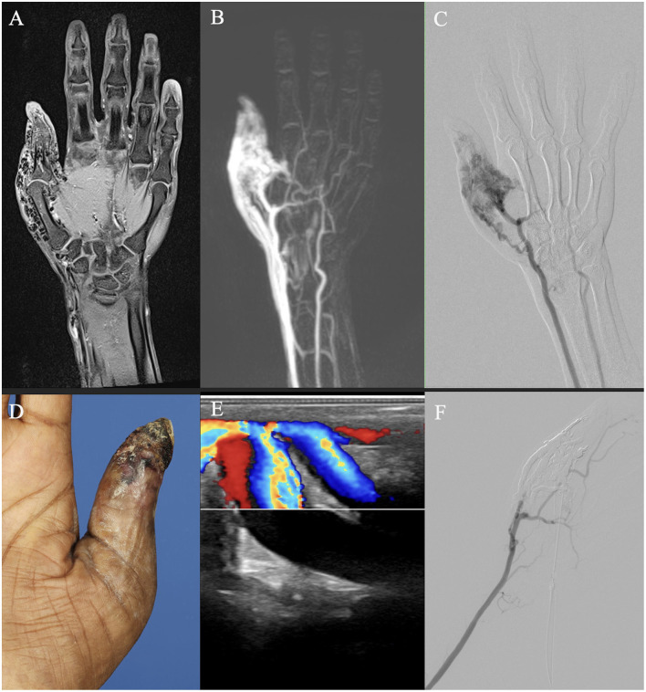
Figure 7.
Imaging of an adult patient with an arteriovenous malformation of the right thumb. (A, B) Magnetic resonance imaging (MRI) shows a dilated vessel with flow voids on a T1 fat-saturated image after intravenous contrast agent admission (A), and a vessel convolute involves the thumb area on a time-resolved angiography with interleaved stochastic trajectories (TWIST) MR angiography with already venous contrast due to the shunt volume (B). (C) Digital subtraction angiography after arterial cubital puncture shows less arterial component than suspected on MRI. (D) Clinical photo of the patient's thumb. (E) Color Doppler ultrasound image demonstrates arterialized flow. (F) DSA after percutaneous embolization demonstrates devascularization of the AVM and earlier arterial perfusion of the palmar arch and the index finger.

