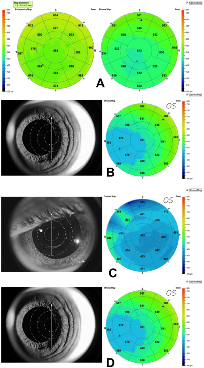
Figure 1.
(A) Corneal Stromal Map (6 mm Ø) generated using Optovue Anterior segment OCT. Notice the non-homogeneous stromal surface as compared to the pachymetry surface. Examples of scans that were excluded. (B) A decentered scan of the left eye caused the corneal apex to shift to the nasal side; (C) Poor scan quality because of scan area coverage; (D) Subepithelial deposits caused by adenoviral keratitis in which the stroma is weakened; if the patient also has corneal ectasia, this condition could cause confusion.

