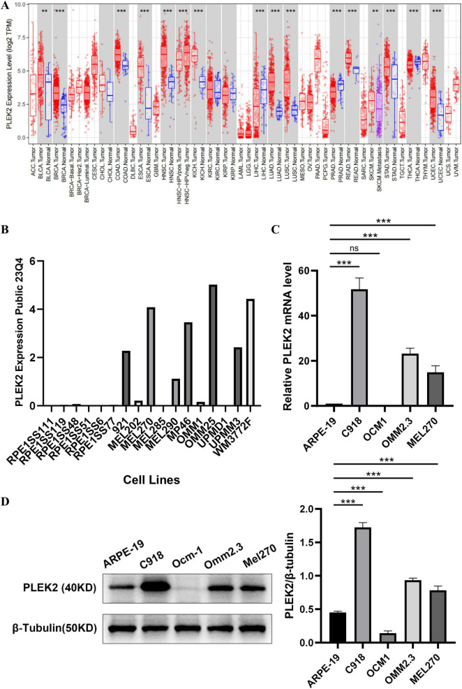
FIGURE 3.
PLEK2 expression was increased in UVM cell lines. (A) PLEK2 mRNA level in various tumor and control tissues. (B) PLEK2 mRNA levels in multiple cell lines. RPE1SS111, RPE1SS119, RPE1SS48, RPE1SS51, RPE1SS6, and RPE1SS77 were retinal pigment epithelial cell lines. 921, MEL202, MEL270, MEL285, MEL290, MP46, OMM1, OMM25, UPMD1, UPMM3 and WM3772F were UVM tumor cell lines. (C) PLEK2 mRNA level was detected by qPCR in ARPE and tumor cell lines, and GAPDH was used as an internal control. (D) PLEK2 protein level was detected by western blot in ARPE and tumor cell lines. Data from at least three independent samples were represented as mean ± SEM; *P < 0.05, **P < 0.01, ***P < 0.001.

