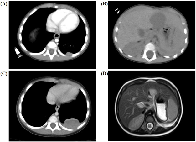
Figure 2.
Secondary chemotherapy and postoperative imaging results for the 6-month-old male infant. (A, B) Abdominal CT showed new space-occupying lesions in the liver (approximately 2.9 cm × 3.0 cm × 2.7 cm) and lung (approximately 1.2 cm × 0.5 cm) after completing 16 weeks of chemotherapy. (C, D) Chest CT and abdominal MRI showed no relief of the multiple new masses and nodules during the whole treatment.

