FIGURE 5.

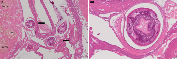
Nematode infection in spotted handfish. (a) Cross sections of nematodes showing round body and external acellular cuticle. Note granuloma forming around the parasites. Some pressure atrophy present (arrows). Pigmented macrophage aggregate (mmc) in the adjacent organ were more numerous than in other unaffected handfish. (b) Eggs present in a cross‐section of a female nematode surrounded by a granuloma.
