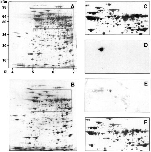
Figure 2.
The effect of reduced NADME activity on mitochondrial protein composition. Total mitochondrial protein from tubers of wild-type and line 35S-ME11 was isolated and separated by two-dimensional SDS PAGE, and stained with silver. The complete gels for wild type and 35S-ME11 are shown in A and B, respectively. The boxes indicate the regions containing NADME subunits that are enlarged in C and F. The gel shown in A was transferred onto nitrocellulose and the resulting western blot probed with antibodies specific to the 59- or 62-kD subunits of NADME. Enlarged regions of these blots are shown in D (62-kD subunit) and E (59-kD subunit). The ovals in C and F indicate the position of the NADME subunits.

