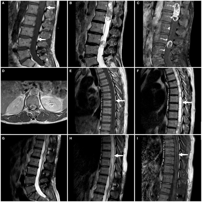
Figure 1.
On the sagittal T1WI sequence, iso-signal occupying lesions (at the white arrow) are discernible separately within the T11-L1 segment and the L3 segment (A). On the sagittal T2WI sequence, both tumors exhibit mixed signals (B). Enhanced T1WI scans in both sagittal and axial planes reveal significant and heterogeneous enhancement of both lesions (C, D). A persistent intramedullary abnormal signal at T4-T10 was seen above the lesion (at the white arrow), showing hypointense on the T1WI and hyperintense on the T2WI (E, F). Follow-up MRI at 3 months confirmed complete tumor removal with no recurrence (G). Sagittal T1- and T2-weighted thoracic MRI images reveal a marked reduction in syringomyelia compared to preoperative findings (white arrow) (H, I).

