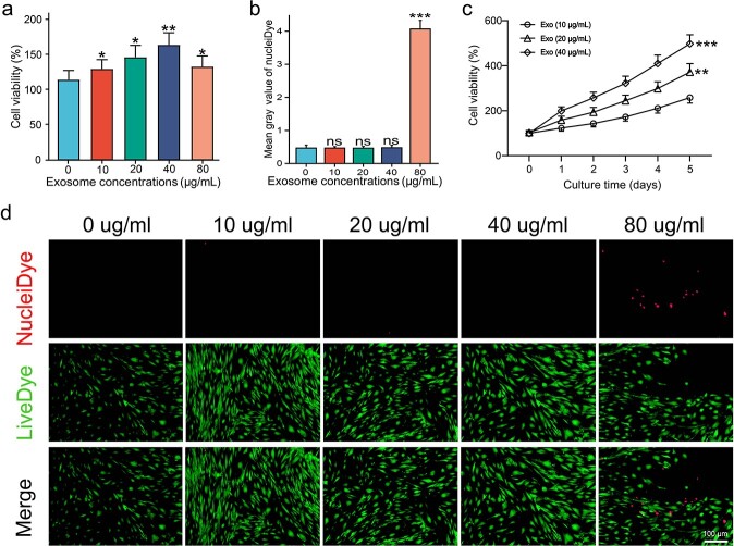
Figure 2.
Optimum ESCs-Exo concentration in HSFBs culture. (a) HSFBs were treated with different concentrations of exosomes for 24 h and cell viability was determined via a CCK8 assay. Scale bar: 50 μm. (b,d) HSFBs were treated with different concentrations of exosomes for 24 h, and cell viability was determined via a live/dead assay. Green, live cells; red, dead cells. Scale bar: 100μm. (c) HSFBs were treated with 20 or 40 μg/ml exosomes for different durations and cell viability was determined via a CCK8 assay. The 0 μg/ml exosome treatment was used as a control. The data are presented as the mean ± SD. *p < 0.05, **p< 0.01, ***p < 0.001 compared with the control; ns, not significant. ESCs pidermal stem cells,HSFBs human skin fibroblasts

