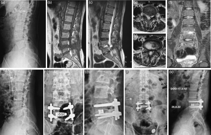
Fig. 1:
Male 22 years, operated micro lumbar discectomy for lumbar disc herniation L4-5. (a) Lateral radiograph before index surgery. (b-f) MRI T1-T2 sagittal, T2 axial and coronal sequences showing intra-discal abscess, left para-discal foraminal abscess, with end-plate destruction. (g) Lateral radiograph showing evident disc space narrowing and irregular end plate. (h, i) Antero-posterior and lateral radiograph showing transforaminal lumbar inter-body fusion executed with bone graft and cage with pedicle screws. (j, k) 95 months final follow-up antero-posterior and lateral radiographs showing stable united reconstruction.

