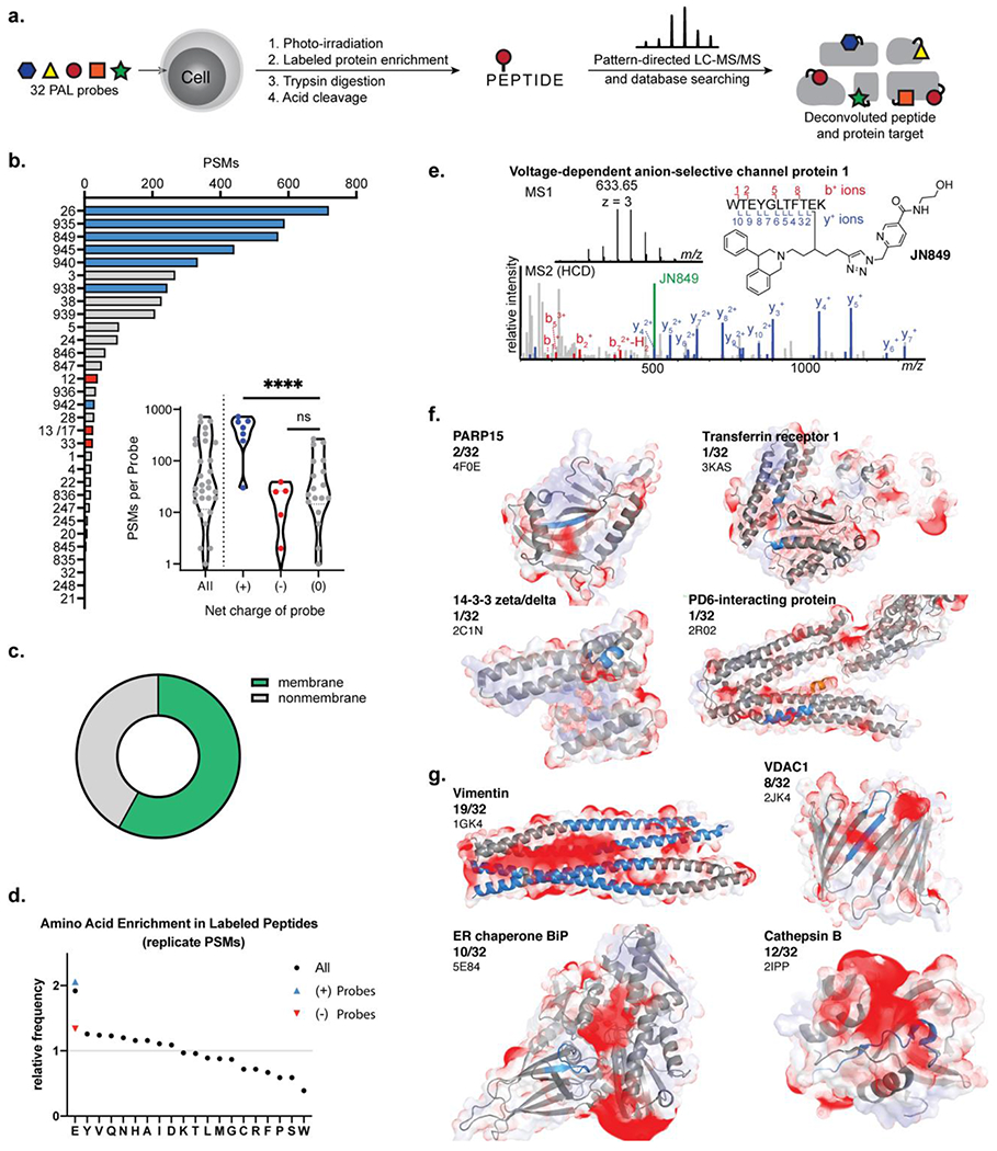
Figure 5.

Binding site mapping with a 32 PAL probe library. (a) Cell cultures were treated simultaneously with 5 or 6 PAL probes which were photo-conjugated to protein binding partners. The conjugated proteins were enriched and the conjugated peptide representing the binding site of the PAL probe was isolated for analysis using isotope-targeted MS. (b) Count of peptide spectral matches (PSMs) assigned to each PAL probe. PAL probes with a net positive charge highlighted in blue, neutral probes in grey, and negatively charged probes in red. (c) Comparison of binding site PSMs from membrane and nonmembrane proteins. (d) Amino acid frequency in the conjugated peptides relative to frequency in human proteome. (e) Annotated mass spectra and assignment of a VDAC1 binding site. (f) Negative electrostatic maps (red) overlaid on the conjugated peptide (blue) for unique binding interactions. (g) Negative electrostatic maps (red) overlaid on conjugated peptide (blue) for frequently conjugated proteins.
