
Figure 7.
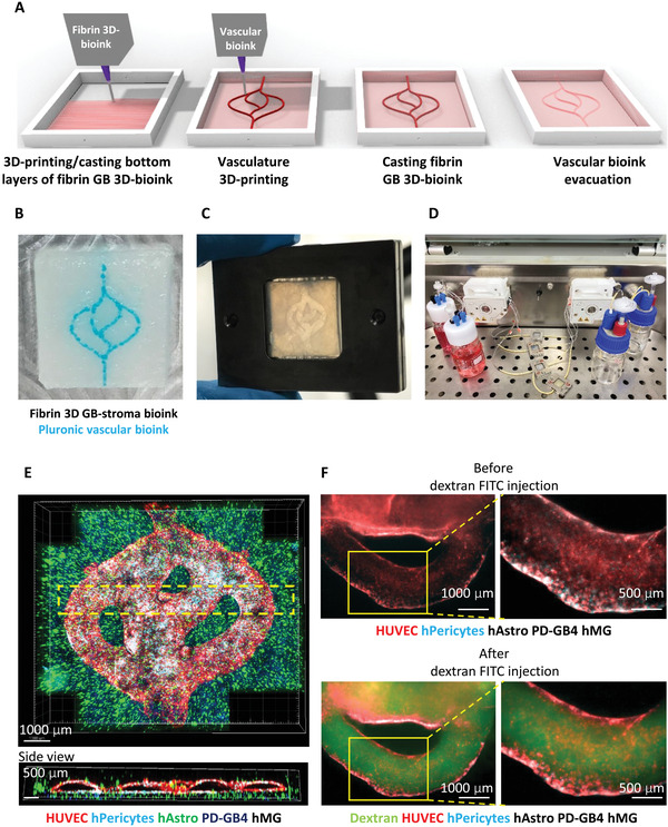
Fibrin brain‐mimicking 3D‐bioink integrated with 3D engineered printed perfusable vascular network. A) Schematic illustration of the 3D‐bioprinting model multistage process. B) 3D‐printed Pluronic‐based vascular bioink (in cyan) on top of 3D‐printed layers of fibrin 3D glioblastoma (GB)‐stroma bioink (in white). C) 3D‐bioprinted vascularized GB model sealed in a metal frame showing the complete perfusion chip. D) The vascularized 3D‐bioprinted GB model is connected to a peristaltic pump through a tubing system, placed in a designated incubator. E) Tiled Z‐stack confocal microscopy images of the 3D‐printed penta‐culture vascularized GB model. Blood vessels are lined with iRFP‐labeled hPericytes (in cyan) together with mCherry‐labeled HUVEC (in red) and surrounded by azurite‐labeled patient‐derived (PD)‐GB4 (in blue), GFP‐labeled human astrocytes (hAstro) (in green), and nonlabeled human microglia (hMG). The dashed box represents a coronal cross‐sectional plane of the vessel. F) Fluorescence microscopy images of the 3D‐bioprinted vascularized GB model before (top) and after (bottom) perfusion of 70‐kDa dextran‐FITC. The 3D‐bioprinted model is composed of a fluorescently labeled vascular network (mCherry‐labeled HUVEC and iRFP‐labeled hPericytes) surrounded by nonlabeled GB‐bioink (hAstro, PD‐GB4, and hMG). Reproduced with permission.[ 121 ] Copyright 2021, American Association for the Advancement of Science (AAAS).