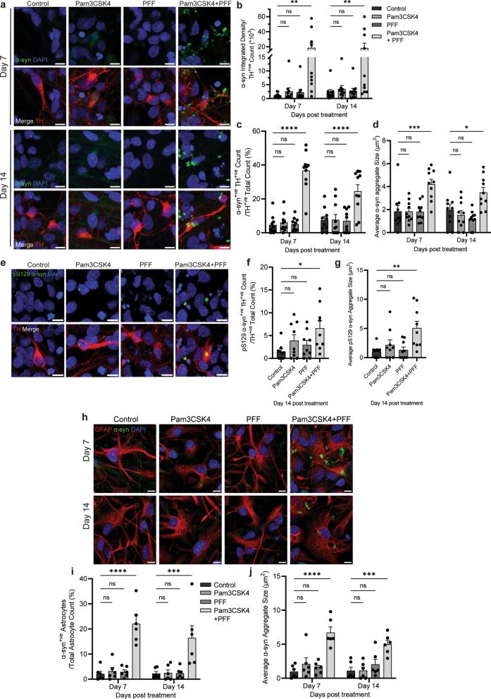
Fig. 4.
TLR2 activation potentiates α-syn PFF mediated pathology in DA neurons and astrocytes. Differentiated PD patient iPSCs were cultured for 7 and 14 days with or without 1 μg/ml Pam3CSK4 and/or 1 μg/ml PFFs. a Cells were fixed at the indicated timepoints and stained for total α-syn (green) and TH (red). Confocal images were taken at 40 × magnification with 6 images captured per condition and used for the analysis of α-syn signal intensity and particle analysis. Scale bar, 10 μm. b Graph shows α-syn integrated density/TH-positive cell number as mean ± SEM (n = 10). c Graph shows the average percentage of TH-positive cells with α-syn aggregates ± SEM (n = 10). d Graph shows α-syn aggregate size (μm2) measured in TH positive neurons displayed as mean ± SEM (n = 10). e Cells were fixed at day 14 post treatment and stained for pS129 α-syn (green) and TH (red). Confocal images were taken at 40 × magnification with 6 images captured per condition and used for the analysis of pS129 α-syn aggregate count and particle analysis in TH positive neurons. Scale bar, 10 μm. f Graph shows the average number of TH positive neurons with pS129 α-syn aggregates ± SEM (n = 9). g Graph shows pS129 α-syn aggregate size (μm2) as mean ± SEM (n = 9). h Cells were fixed at the indicated timepoints and stained for total α-syn (green) and GFAP (red). Confocal images were taken at 40 × magnification with 6 images captured per condition and used for the analysis of α-syn aggregate count and particle analysis. Scale bar, 10 μm. i Graph shows average percentage of GFAP positive cells with α-syn aggregates ± SEM (n = 6). j Graph shows α-syn aggregate size (μm2) in astrocytes as mean ± SEM (n = 6). For all graphs *P < 0.05, **P < 0.01, ***P < 0.001, ****P < 0.0001, ns = not significant

