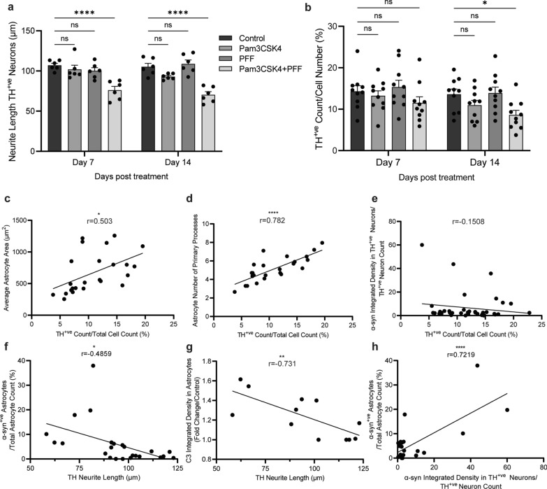
Fig. 9.
Specific loss of DA neurons with Pam3CSK4 plus α-syn PFF treatment. Differentiated midbrain cells derived from PD patient iPSCs were cultured for 7 and 14 days with 1 μg/ml Pam3CSK4 and/or 1 μg/ml PFFs, or medium only for the untreated control. a Graph shows the length of neurites extending from the soma of TH-positive neurons (μm) as mean ± SEM. 30 cells in each replicate were included in the analysis (n = 6). b Graph shows the number of TH positive neurons/DAPI (%) displayed as mean ± SEM (n = 10). c–h Graphs show Pearson correlation between two variables, as described on the axis. For all graphs *P < 0.05, **P < 0.01, ****P < 0.0001, ns = not significant

