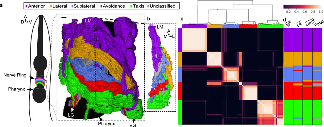
Fig. 1. Five densely connected neurite clusters comprise the nerve ring neuropil.
a, The nerve ring neuropil (<4% of the worm’s body length and most synaptically dense region of the nervous system) includes neurites of 181 L4 (185 adult ) neurons. Complete volumetric reconstruction of the L4 neuropil spans 36 μm (Supplementary Video 3). 15 μm -long region (inset): left view, superficial neurons removed. D: dorsal, V: ventral, A: anterior. b, A 250 nm oblique volumetric slice at approximately the lateral midline (LM) rendered with no processes removed (right). A/P: anterior/posterior, M/L: medial/lateral, LG/VG: lateral/ventral ganglia. Scale bar: 1 μm Neurites with relatively high spatial affinity (but no physical boundaries) form spatially ordered clusters along anterior-posterior axis. c, Cluster matrix: frequency that cells and cluster together across the population : Row and column order minimized frequency variance along the diagonal. Clusters were then ordered to visually match AP ordering (original ordering in Extended Data Fig. 5i). Top: Dendrogram of the hierarchical clustering. d, Clustering results of model , and populations (Extended Data Fig. 5i) and consensus cluster assignment across the 3 populations. Row and column order same as rows in c. 7 cell classes (ADE, ALN, AVA, RID, RIR, RMD, URX) with discrepant cluster assignments among the 3 populations are unclassified (gray). perturbed datasets per population (Methods).

