. 2024 Dec 13;12:RP90419. doi: 10.7554/eLife.90419

Figure 2. Loss of Slc39a5 results in elevated circulating and hepatic zinc levels in mice.

Serum zinc (A) and hepatic zinc (B) in Slc39a5+/+, Slc39a5-/-, and Slc39a5+/-mice at 40 wk of age, n=16–18. **p<0.01, ***p<0.001, two-way ANOVA with post hoc Tukey’s test.

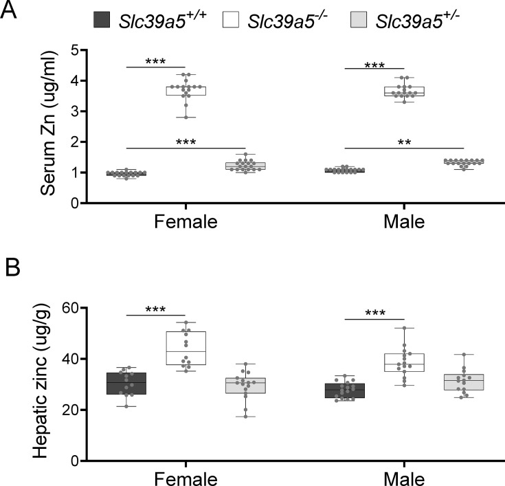
Figure 2.

Figure 2—figure supplement 1. Generation and characterization of the Slc39a5-/- mice.

Figure 2—figure supplement 1.

(A) Schematic representation of the Slc39a5 null allele. (B) Slc39a5 gene expression in liver and duodenum of Slc39a5-/- mice at 20 wk of age, n=3–6. (C) Immunoblotting analyses demonstrating an absence of SLC39A5 protein in the liver of Slc39a5-/- mice at 34 wk of age. (D–E) Heterozygous loss of Slc39a5 does not reduce fasting blood glucose in (D) Lepr-/- mice (at 20 wk of age) and in (E) mice challenged with high-fat high fructose diet (HFFD) for 18 wk. *p<0.05, **p<0.01, ***p<0.001, two-way ANOVA with post hoc Tukey’s test.
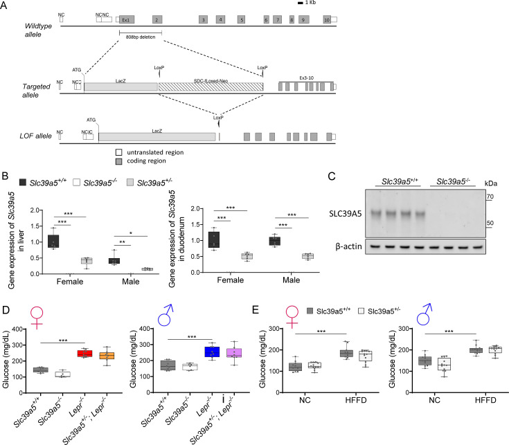
Figure 2—figure supplement 1—source data 1. Original files of the full raw uncropped, unedited blots.
Figure 2—figure supplement 1—source data 2. Figures with the uncropped blots with the relevant bands clearly labelled.
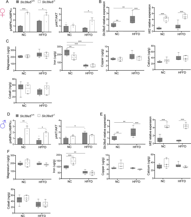
Figure 2—figure supplement 2. Loss of Slc39a5 does not alter hepatic magnesium, iron, copper, calcium, and cobalt levels in mice challenged with high-fat high fructose diet (HFFD).

Figure 2—figure supplement 2.

Female (A–C) and male (D–F) mice were fed HFFD or NC for 30 wk. (A, D) Densitometric analysis of hepatic AMPK and AKT. (B, E) Hepatic gene expression of Slc39a5 and Mt2. (C, F) Hepatic ion quantification by flame atomic absorption spectrometry. *p<0.05, **p<0.01, ***p<0.001, two-way ANOVA with post hoc Tukey’s test.