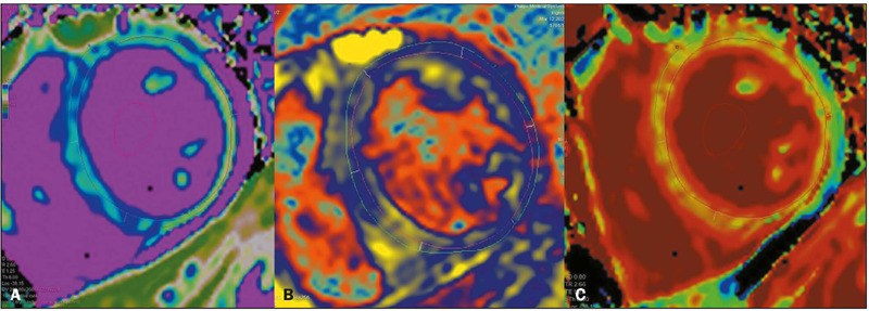
Figure 15.

A 29-year-old patient with acute dengue-induced myocarditis. MRI with native T1 mapping of myocardial tissue (A), T2 mapping of myocardial tissue (B), and depiction of myocardial extracellular volume (C), showing diffuse signal alteration, indicating a myocardial inflammatory process. (Images kindly provided by Dr. Joalbo Matos de Andrade)..
