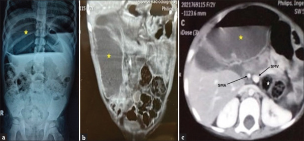
Figure 1.
(a) Erect abdominal X-ray showing a grossly dilated bowel loop occupying almost 50% of the abdomen (yellow star) (b) CT angioenterography of abdomen (coronal section) showing a grossly dilated small bowel loop of diameter 5.2 cm (yellow star) (c) CT angioenterography of the abdomen (axial section) showing reversal of SMA and SMV relationship, with cecum and transverse colon (white star) lying posterior to the small bowel (yellow star), suggestive of reversed intestinal rotation

