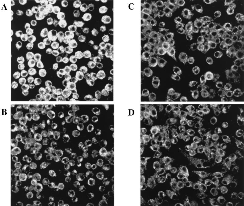
FIG. 3.
J774A.1 cells were grown on Thermonox coverslips; TfRs were labeled with an anti-TfR monoclonal antibody and examined by confocal microscopy for TfR expression. (A) J774A.1 cells infected for 16 h with viable C. burnetii show increased TfR expression as well as a rounding up of infected cells. (B) J774A.1 cells incubated in conditioned medium from persistently infected J774A.1 cells. These cells show no upregulation of TfR but do exhibit the rounding up seen in infected cells. (C) J774A.1 cells inoculated with inactivated C. burnetii and (D) uninoculated J774A.1 cells exhibit a basal level of TfR expression and no morphological changes.

