Abstract
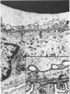
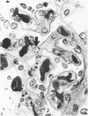
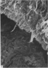
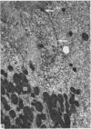
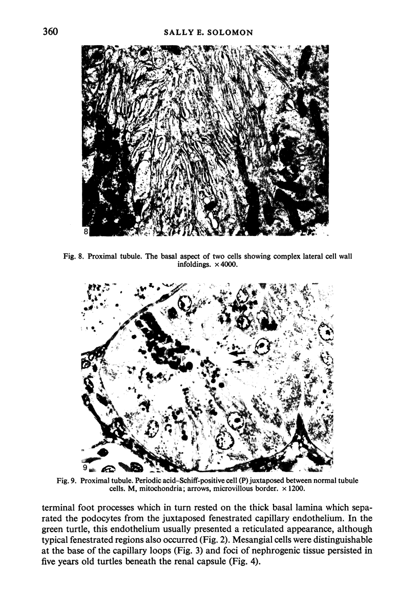
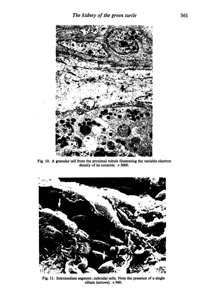
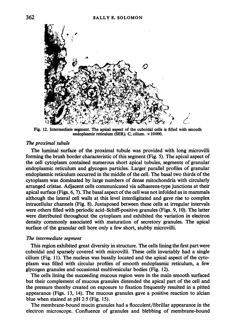
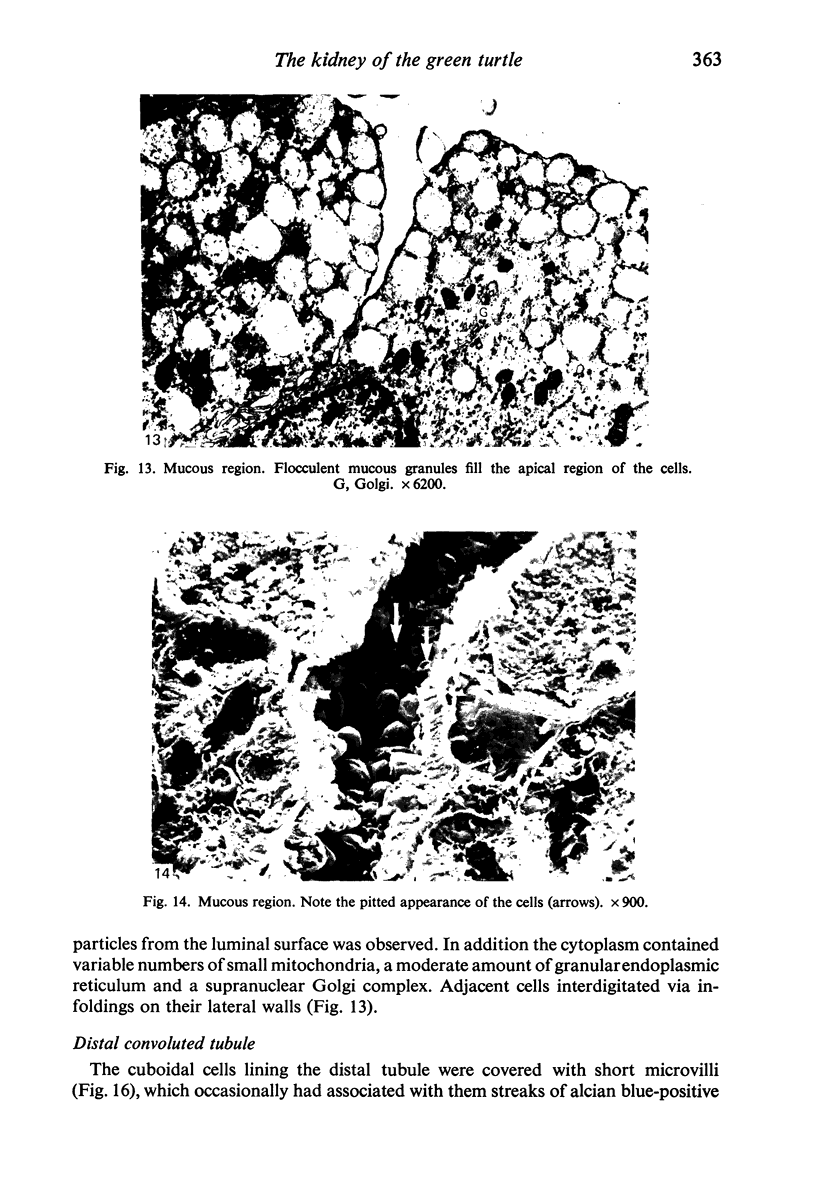
The kidneys of the green turtle are flattened, lobed and closely applied to the posterior wall of the pleuroperitoneal cavity. The ability to differentiate new functional nephrons is retained by five years old animals. The functional nephron comprises a glomerulus, proximal tubule, intermediate segment which can be subdivided into a proximal non-secretory segment and a distal mucus secreting segment, distal convoluted tubule and collecting tubule. Both the proximal and distal tubules exhibit complex foldings on their lateral cell walls in contradistinction to the characteristic basal infoldings observed in mammalian tubules.
Full text
PDF
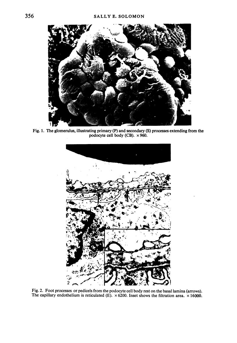
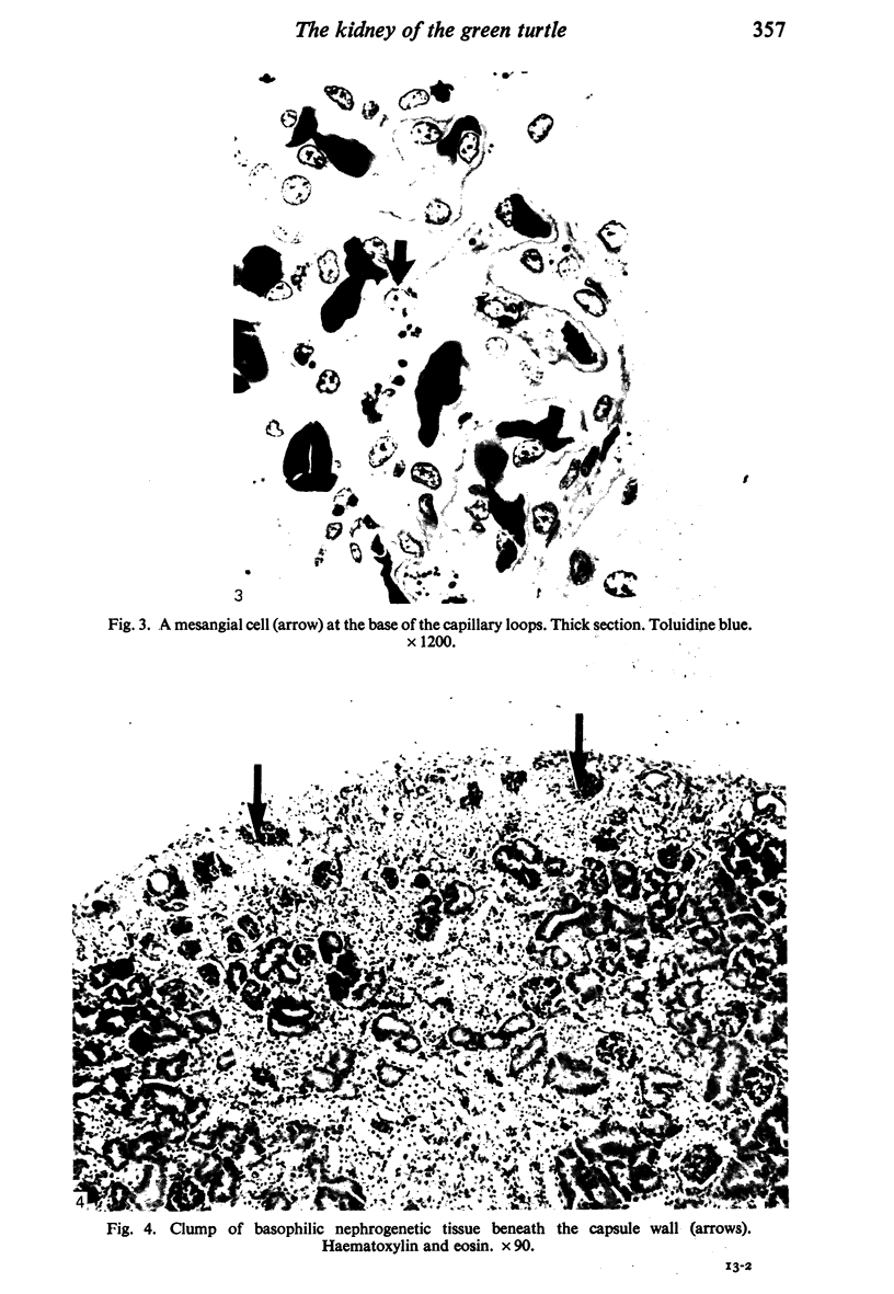
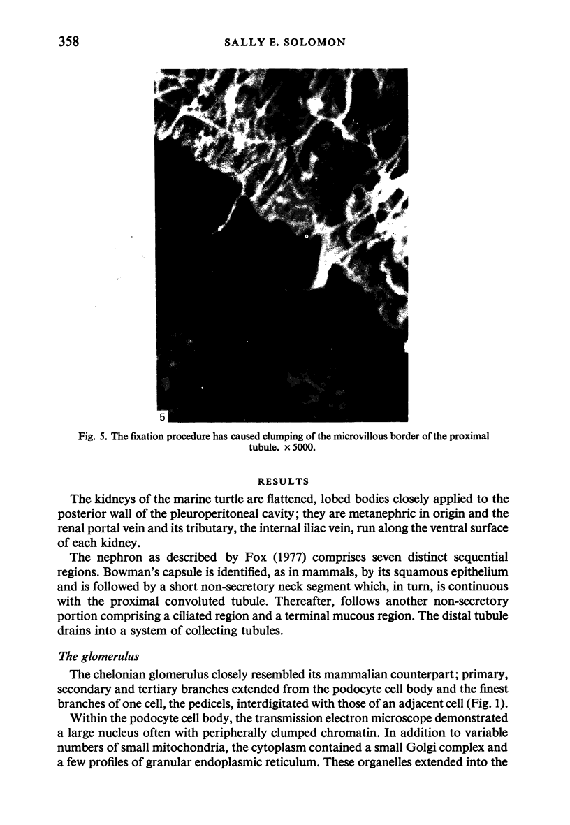
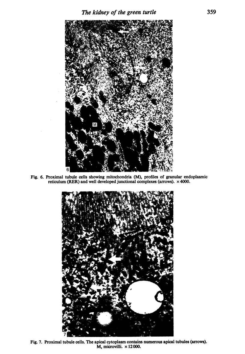
Images in this article
Selected References
These references are in PubMed. This may not be the complete list of references from this article.
- Davis L. E., Schmidt-Nielsen B. Ultrastructure of the crocodile kidney (Crocodylus acutus) with special reference to electrolyte and fluid transport. J Morphol. 1967 Apr;121(4):255–276. doi: 10.1002/jmor.1051210402. [DOI] [PubMed] [Google Scholar]
- Farquhar M. G., Palade G. E. Adenosine triphosphatase localization in amphibian epidermis. J Cell Biol. 1966 Aug;30(2):359–379. doi: 10.1083/jcb.30.2.359. [DOI] [PMC free article] [PubMed] [Google Scholar]
- Kelly D. E. The Leydig cell in larval amphibian epidermis. Fine structure and function. Anat Rec. 1966 Mar;154(3):685–699. doi: 10.1002/ar.1091540314. [DOI] [PubMed] [Google Scholar]
- Lane B. P., Europa D. L. Differential staining of ultrathin sections of Epon-embedded tissues for light microscopy. J Histochem Cytochem. 1965 Sep-Oct;13(7):579–582. doi: 10.1177/13.7.579. [DOI] [PubMed] [Google Scholar]
- Maunsbach A. B. Observations on the ultrastructure and acid phosphatase activity of the cytoplasmic bodies in rat kidney proximal tubule cells. With a comment on their classification. J Ultrastruct Res. 1966 Oct;16(3):197–238. doi: 10.1016/s0022-5320(66)80059-x. [DOI] [PubMed] [Google Scholar]
- Rothwell B., Solomon S. E. The ultrastructure of the follicle wall of the domestic fowl during the phase of rapid growth. Br Poult Sci. 1977 Sep;18(5):605–610. doi: 10.1080/00071667708416409. [DOI] [PubMed] [Google Scholar]
- Tessenow W. Die chemische Zusammensetzung und die Bedeutung der PAS-positiven Granula in der supranucleären Zone der Nierentubuluszellen bei Xenopus laevis Daud. Acta Histochem. 1965 Aug 14;21(5):313–322. [PubMed] [Google Scholar]


