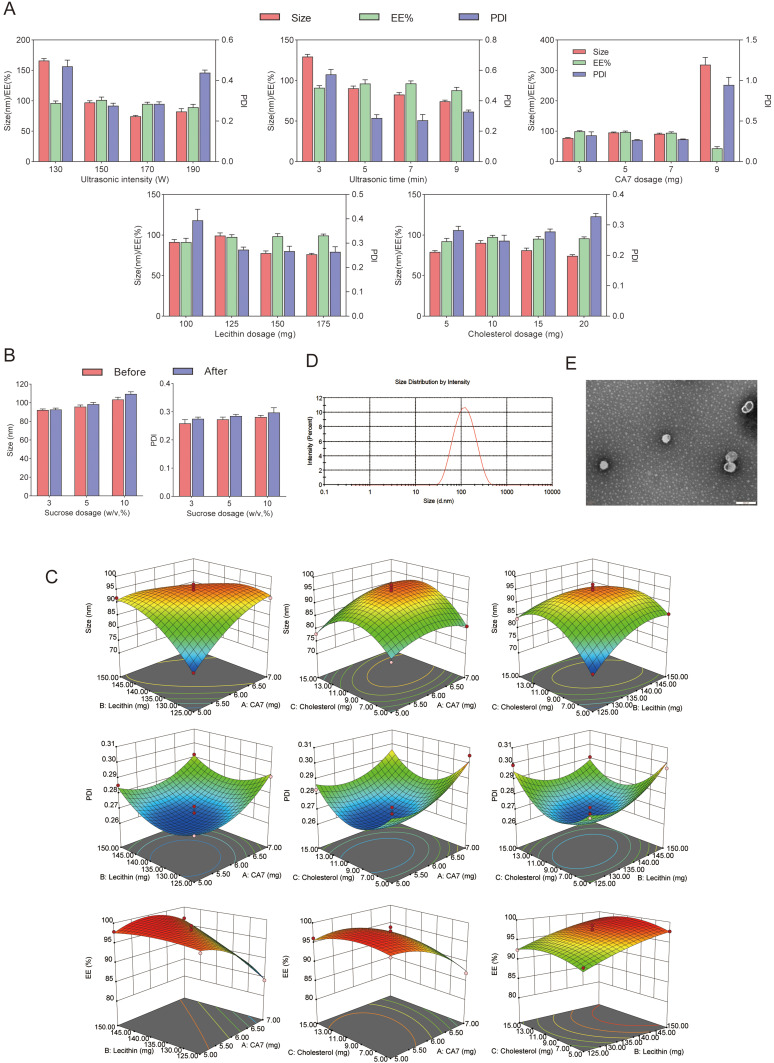
Figure 2.
(A) The effects of ultrasound intensity, ultrasound time, CA7 dosage, lecithin dosage, and cholesterol dosage on PS, PDI, and EE of CA7-LP. (B) The effects of lyophilized protectants on PS and PDI of CA7-LP. (C) Two-way interaction contour plots of three factors. (D) PS distribution of CA7-LP. (E) Morphology of CA7-LP by transmission electron microscopy.

