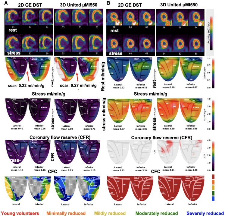
Figure 4.
3D United µMI550 compared with established 2D GE DST for extremes of myocardial perfusion. (A) A 63-year-old male with known lateral-inferior myocardial infarction (MI), coronary artery bypass surgery (CABG), and PCI with reduced ejection fraction of 35%. Perfusion at rest in the outlined territory of transmural scar (white line) is 0.22 mL/min/g on the 2D DST and 0.27 on the United, respectively. (B) A 30-year-old male participant without risk factors or a family history of heart disease shows reproducibility between 2D and 3D PET-CT of rest and stress millilitres per minute per gram and CFR > 4 cc/min/g.

