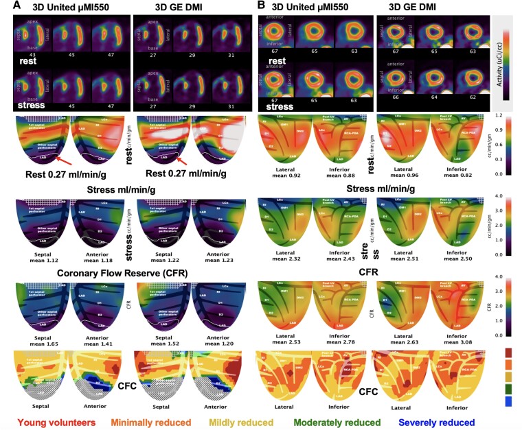
Figure 5.
Serial images of two participants on different days on the 3D GE DMI and 3D United µMI550 for extremes of CAD severity. (A) The iso-contour of the fixed perfusion defect is identical at 0.27 mL/min/g for both scanners and within the acceptable range of MBF for transmural myocardial scar (17). (B) Participant with comparable perfusion, CFR, and CFC on each 3D system.

