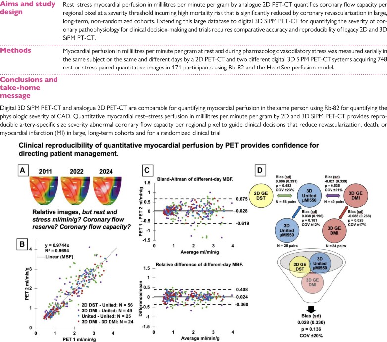
Structured Graphical Abstract.
Rationale and summary of the study. In large non-randomized patient cohorts and in a randomized trial followed over 14 years, CFC by 2D PET-CT quantifies CAD severity favouring lifestyle-medical treatment while reducing coronary interventions to physiologically severe, high-risk, obstructive CAD having survival benefit after revascularization. Documenting equivalency, reproducibility, and precision of high-sensitivity 3D SiPM PET-CT compared with 2D PET-CT systems extends this extensive knowledge base derived from 2D PET-CT using Rb-82. The addition of 3D SiPM PET-CT with optimal acquisition–reconstruction protocols and perfusion models provides confidence to physicians and patients in quantifying physiologic CAD severity to guide personalized management and randomized trials of CAD management. For a patient with serial scans over 13 years (A), relative stress images are unchanged on 2D GE DST, 3D United µMI550, and 3D GE DMI PET-CT without quantitative perfusion defining the status of severe CAD. Subgroup perfusion correlation in millilitres per minute per gram (B) among all perfusion measurements with Bland–Altman plots (C) of perfusion difference and relative difference (difference/mean) is the evidence for their combined comparison (D). Serial 154 rest–stress millilitres per minute per gram paired measurements on different days in the same person between analogue 2D GE DST, the 3D United µMI550, and the 3D GE DMI PET-CT systems produce the largest comparison to date of ‘different-day’ perfusion (B, C, D). Mean COV is ±20% for methodological plus day-to-different-day biological variability, providing confidence to physicians and patients for quantifying physiologic CAD severity to guide personalized management or randomized trials (D).

