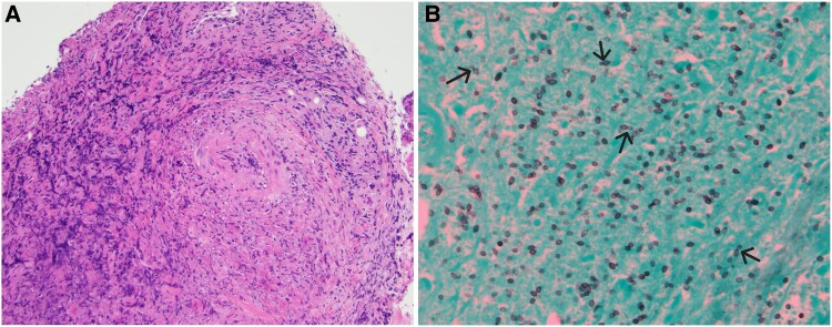
Figure 1.
Supraglottic biopsy. (A) The image shows ulceration with loss of surface epithelium. Beneath is marked acute and chronic inflammation with necrotic cellular debris throughout the biopsy. Perivascular inflammation is prominent. No definitive granulomas are identified. The absence of granuloma formation may be attributed to the patient's use of a tumor necrosis factor-alfa inhibitor (hematoxylin and eosin, 10×). (B) Gomori-methanamine silver stain highlights abundant small oval yeast forms with occasional narrow-based budding. No pseudohyphae are identified. The histologic differential was narrowed to histoplasmosis, cryptococcosis, and emergomycosis. The mucicarmine stain was negative, which ruled out classic Cryptococcus. The clinical picture was not consistent with emergomycosis. ©2024 Ellen Giampoli

