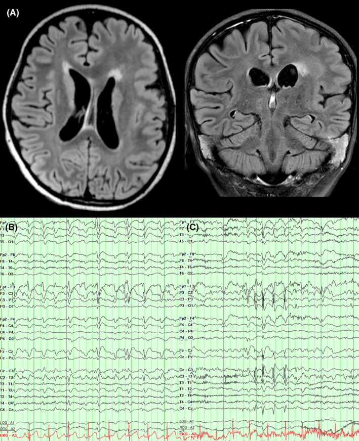
Figure 1.

Baseline structural and electrographic findings. (A) Axial and coronal T2 FLAIR images demonstrating diffuse left frontal pachygyria and polymicrogyria. (B) EEG recording 2 months prior to status presentation revealed abundant left frontopolar epileptiform discharges that were continuous at 2 Hz during non‐rapid eye movement sleep. (C) EEG recording 2 months prior to status presentation also revealed bursts of left central fast activity and spikes that coincided with right hand and face myoclonic seizures.
