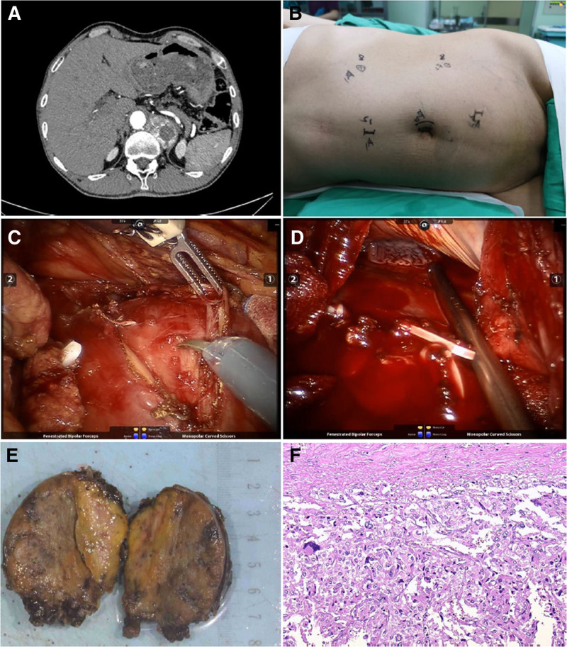
Figure 1.
Radiological, surgical procedures, and pathologic diagnosis in Case A. (A) The CT showed an ovoid mass in the plane of the area between T11 and T12 vertebrae, within the left septum of the spine. (B) The observation mirror was positioned on the left side next to the umbilicus, operating arm No. 2 was placed 2 cm below the rib margin in the midclavicular line, operating arm No. 1 was placed 10 cm above the anterior superior iliac spine, the assistant’s channel was placed 5 cm away from the umbilicus to the lateral side of the pubic symphysis, and the alternate assistant’s channel was placed 10 cm above the umbilicus. (C) Incision of the diaphragm to expose the tumor. (D) Diaphragm rupture. (E) Surgical specimen of the ectopic pheochromocytoma. (F) Pathological findings of the resected specimen: Haematoxylin and eosin (HE) staining lower-power field. CT = computed tomography, HE = haematoxylin and eosin.

