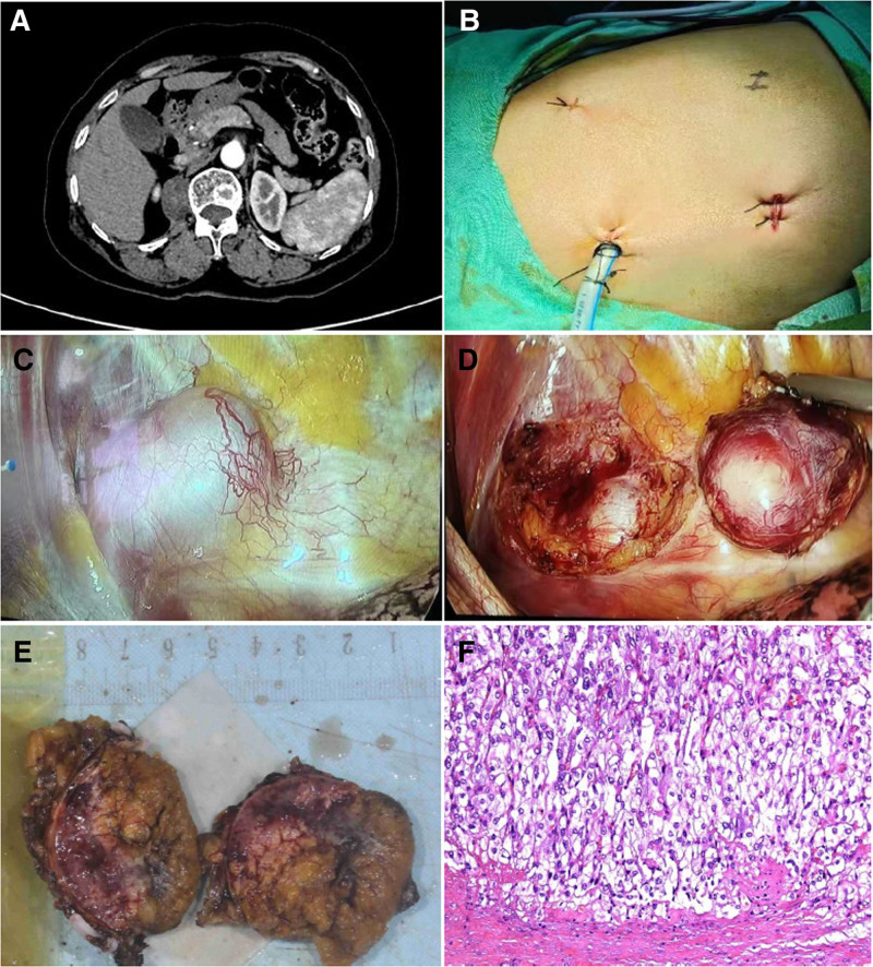
Figure 2.
Radiological, surgical procedures, and pathologic diagnosis in Case B. (A) The CT showed a roundish slightly hypodense lesion adjacent to the spine in the right diaphragmatic angle. (B) The main surgical incision is 4th intercostal space along the midaxillary line; the obersvation hole is 7th intercostal space along the midaxillary line, and the auxiliary incision is 9th intercostal space along the subscapular line. (C) Exposure of the tumor. (D) Remove the tumor. (E) Surgical specimen of the ectopic pheochromocytoma. (F) Pathological findings of the resected specimen: Haematoxylin and eosin (HE) staining lower-power field. CT = computed tomography, HE = haematoxylin and eosin.

