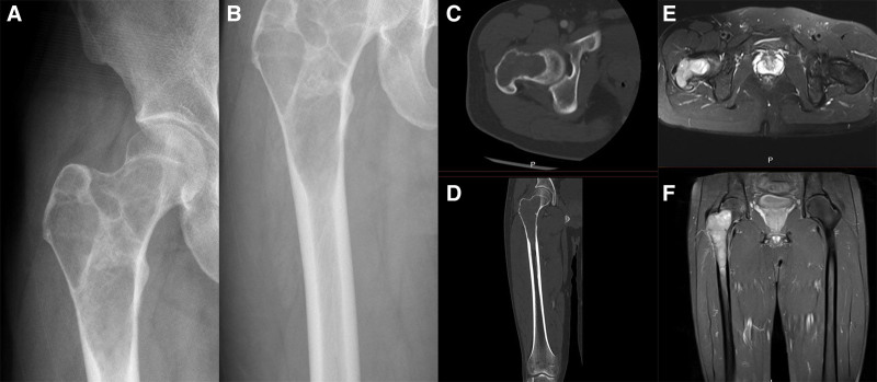
Figure 1.
(A and B) Preoperative anteroposterior radiographs showed extensive cystic lesions in the subtrochanter, neck, and intertrochanter of the femur. (C and D) Preoperative computer-enhanced tomography showed no pathological fracture of the proximal femur, and the cortical bone was damaged but intact. (E and F) Preoperative magnetic resonance imaging showed that the lesion did not infiltrate the surrounding soft tissue.

