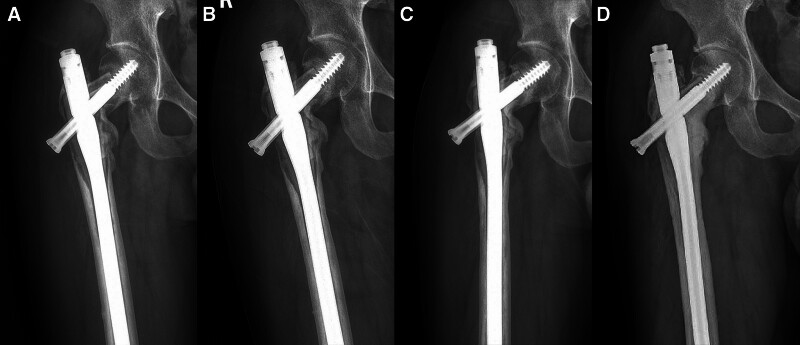
Figure 3.
(A) Postoperative anteroposterior radiographs of the proximal femur showed bone resorption and atrophy at 3 months. (B) After 7 months of follow-up, the intramedullary nail showed signs of loosening. (C) After 12 months of follow-up, bone grafting began to heal and internal fixation gradually stabilized. (D) After 40 months of follow-up, the bone defect was healed and the intramedullary nail was stable in place.

