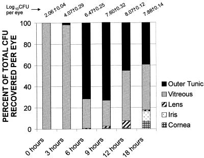
FIG. 2.
Ocular tissue distribution of B. cereus during experimental endophthalmitis. Ocular tissues (cornea, iris, lens, vitreous, and outer tunic) were recovered at various times postinfection for quantification of B. cereus. The charted values represent the percentage of organisms recovered from each tissue compared with the cumulative number of organisms recovered per eye (listed at the top; data from Fig. 1A). Because E. faecalis and S. aureus were recovered exclusively from posterior segment tissues during all stages of infection, those data are not included.

