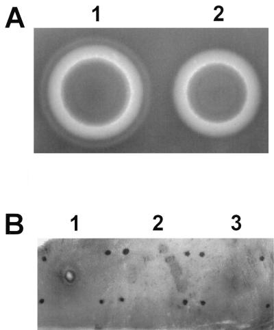
FIG. 3.
Phenotypic analysis of wild-type and hemolysin BL-deficient strains. (A) Single colonies of MGBC145 (plate 1) and CJ145-1.1 (plate 2) were isolated on 2.5% sheep erythrocyte agar. Presence of the discontinuous zone of hemolysis (as seen in plate 1) indicated hemolysin BL activity. (B) Concentrated supernatants of MGBC145 (sample 1), uninoculated BHI (sample 2), or CJ145-1.1 (sample 3) were injected intradermally. Presence of a necrotic center surrounded by a blue (dark) zone (as seen in sample 1) indicated hemolysin BL activity.

