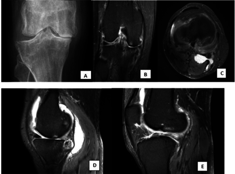
Fig. 3.

(Representative case). A 39-year-old woman presented with bilateral knee pain for the past 2 to 3 years. Her Western Ontario and McMaster scoring system (WOMAC) total pain score was 12/20. ( A ) Posteroanterior (PA) X-ray shows severe reduction of medial joint space with large osteophytes, subchondral sclerosis, and subchondral cysts suggesting Kellgren–Lawrence grading system (K-L) grade 4. ( B ) Coronal proton density fat-saturated (PDFS) image shows full-thickness cartilage loss in the medial femoral and tibial articular surface with large osteophytes and severe narrowing of the joint space. ( C ) Axial PDFS image shows Baker's cyst between the tendons of semimembranosus and medial head of the gastrocnemius. ( D ) Sagittal PDFS image shows complete maceration of the posterior horn of the medial meniscus. ( E ) Sagittal PDFS image reveals complete anterior cruciate ligament (ACL) tear.
