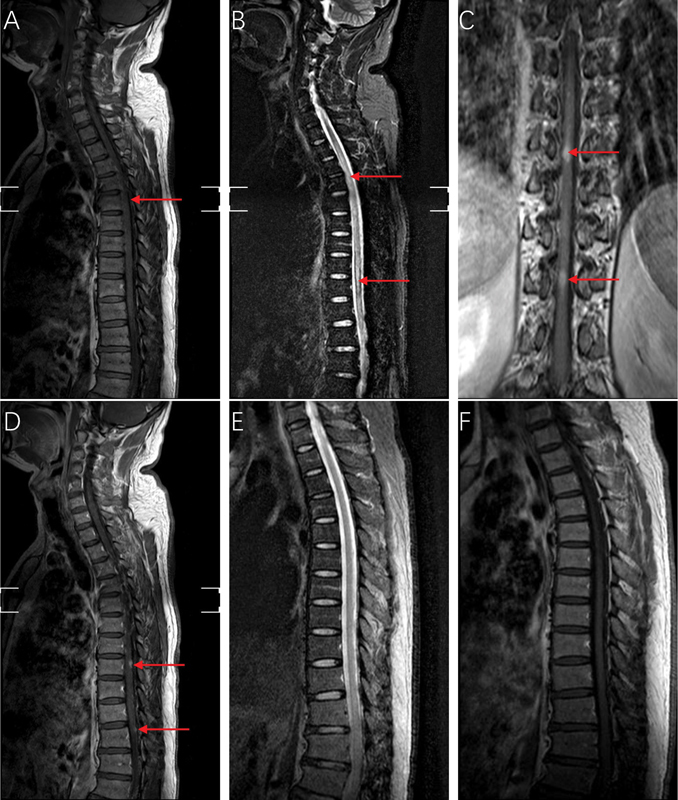
Fig. 1.

Magnetic resonance imaging of the spinal cord of the patient. ( A ) Sagittal T1-weighted image of the spinal cord showing spinal cord swelling, ( B ) sagittal T2-weighted image of the spinal cord showing long segmental intramedullary hyperintensity, ( C ) coronal T1-weighted enhanced image of the spinal cord showing two enhanced lesions, ( D ) sagittal T1-enhanced image of the spinal cord showing two enhanced lesions (candle guttering appearance), ( E ) sagittal T2-weighted image of the spinal cord after treatment indicating complete resolution of the spinal cord lesions, and ( F ) sagittal T1-weighted image of the spinal cord after treatment indicating complete resolution of the spinal cord swelling.
