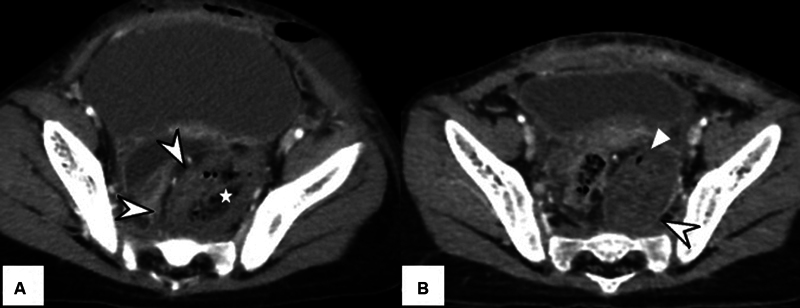
Fig. 3.

( A ) Axial computed tomography (CT) section through the pelvis on the third postoperative day showed a lentiform well-defined low-density collection containing air foci (*) located within a hyperdense hematoma in the left side of the pelvis ( arrowheads ). ( B ) Repeat imaging demonstrates a persistent hematoma. However, the component containing air foci had almost completely resolved.
