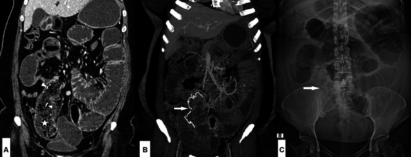
Fig. 6.

A 35-year-old male patient with a history of previous surgery presented with acute abdomen. ( A ) Coronal contrast-enhanced computed tomography ((CECT) image showed dilated small bowel loops with the transition zone in the right lower abdomen and the distal ileal loops collapsed. The small bowel proximal to the transition zone had mottled air lucencies (*) and few radiodense foci ( arrowheads ). ( B ) Thick slab reconstruction revealed a thin serpiginous radiodensity within the bowel lumen ( arrow ). ( C ) Supine abdominal radiograph showed a similar radiodensity in the right lower abdomen ( arrow ). These findings were consistent with intraluminal gossypiboma in the mid-ileum with small bowel obstruction.
