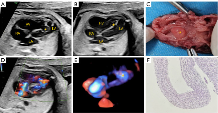
Figure 1.
Prenatal image of VSA. (A) Early systole: aneurysm bulges from the body of the right ventricle into the left ventricle. (B) Late systole: the aneurysm bulges towards the right ventricle. (C) Color Doppler demonstrating the aneurysm. (D) The imaging of aneurysm with STIC. (E) Autopsy specimen. (F) Pathological manifestations of aneurysms (hematoxylin and eosin stain, 100×). The asterisk (*) indicates the VSA. RA, right atrium; RV, right ventricle; LA, left atrium; LV, left ventricle; VSA, ventricular septal aneurysm; STIC, spatiotemporal image correlation.

