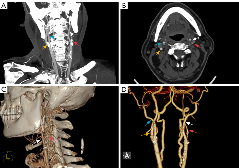
Figure 3.
MPR images (A,B) and VR reconstruction images (C,D) of a case with left-sided carotid near-occlusion with full collapse. The left ICA is severely stenotic at its origin, and the distal lumen is completely collapsed, “thread-like” (red arrow), and smaller than the contralateral ICA (yellow arrow) and the ipsilateral ECA (white arrow), while the right ICA (yellow arrow) is thicker than is the ipsilateral ECA (light blue arrow). MPR, multiplanar reconstruction; VR, volume rendering; ICA, internal carotid artery; ECA, external carotid artery.

