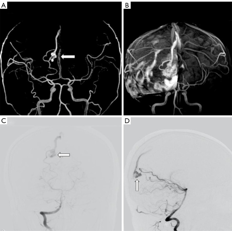
Figure 2.
Effects of new hemorrhage on image quality using the two MRA techniques. Images of right occipital lobe BAVM in a 26-year-old man with acute intracranial hemorrhage and diffused subdural hematoma. (A) The feeding artery, nidus, and the draining vein were visible on silent MRA (arrow). The image quality scores are as follows: feeding artery: 3; nidus: 3, and draining vein: 3. (B) TOF MRA exhibited diffused hyperintensity. The feeding artery was invisible, and the nidus and the draining vein were indistinct. The image quality scores are as follows: feeding artery: 0; nidus: 1; and draining vein: 1. (C,D) The front and lateral view of the right vertebral artery angiography showed a small nidus fed by the right posterior cerebral artery (arrows). BAVM, brain arteriovenous malformation; MRA, magnetic resonance angiography; TOF MRA, time-of-flight MRA.

