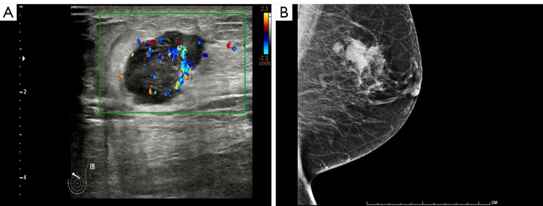
Figure 1.
Imaging examination of the left breast. (A) Ultrasound examination revealed an irregular hypoechoic mass in the left breast with well-defined borders and abundant blood flow signals, measuring approximately 2.6 cm × 1.9 cm × 2.2 cm. No suspicious axillary lymph nodes were found. (B) Mammography showed a high-density mass shadow in the left breast, with unclear boundaries and irregular shape, measuring approximately 3.1 cm × 2.3 cm, with distorted surrounding glandular structures. The left breast also showed orbit-like calcification.

