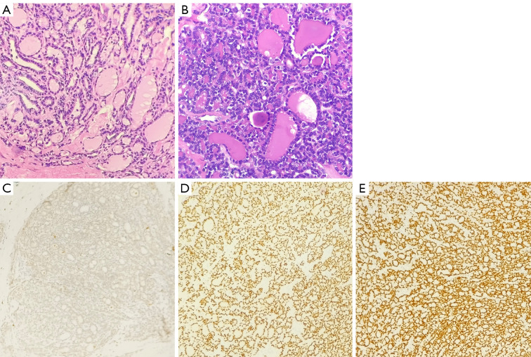
Figure 3.
Histopathological and immunohistochemical features of the primary and metastatic lesions. (A) H&E stain (×200) of the left thyroid lobe showing FTC with colloid-filled follicles. (B) H&E stain (×200) of the left breast mass revealing a tumor composed of thyroid follicles filled with colloid, without a residual breast tissue, indicating metastatic rather than primary breast carcinoma. (C) GATA-3 immunostaining (×100) showing negative nuclear expression in the breast metastasis, supporting non-mammary origin. (D) PAX-8 immunostaining (×100) showing positive nuclear expression in the breast metastasis, confirming thyroidal origin. (E) TTF-1 immunostaining (×100) showing positive nuclear expression in the breast metastasis, further affirming its thyroidal derivation. H&E, hematoxylin and eosin; FTC, follicular thyroid carcinoma; GATA-3, GATA binding protein 3; PAX-8, paired box gene 8; TTF-1, thyroid transcription factor-1.

