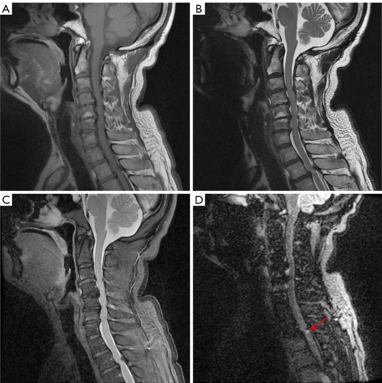
Figure 1.
Sagittal imaging of the spine. (A) Sagittal T1WI. (B) Sagittal T2WI. (C) Sagittal T2WI with fat-saturated series. (D) DIR. Spine magnetic resonance imaging revealed multilevel disc herniation, the DIR sequence showed abnormal signals at the posterior cord of the second thoracic vertebra to the third thoracic vertebra (T2–3) (long red arrow). T1WI, T1-weighted image; T2WI, T2-weighted image; DIR, double inversion recovery.

