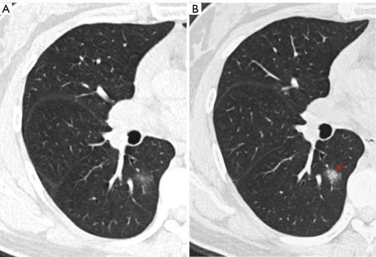
Figure 4.
A 52-year-old female with an incidental GGN. (A) An axial CT image showed a 14-mm round and ill-defined pGGN with a mean density of −609 HU located in the right lower lobe. (B) A follow-up CT scan performed 27 days later showed an increase in density (−324 HU) and the development of new solid components (red arrow). The histopathologic analysis revealed fibrous tissue proliferation with less inflammatory cell infiltration. GGN, ground-glass nodule; CT, computed tomography; pGGN, pure ground-glass nodule; HU, Hounsfield unit.

