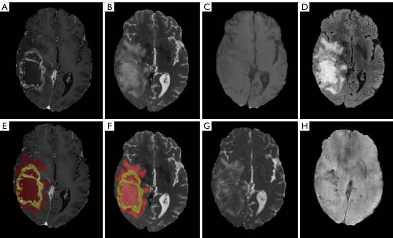
Figure 2.
Example for MRI images of a patient with glioblastoma, IDH wild type, WHO Grade 4, MGMT unmethylated: T1CE (A), T2WI (B), T1WI (C), FLAIR (D), segmentation on T1CE (E), segmentation on T2WI (F), ADC (G), and SWI (H). Yellow area: enhancing tumor area. Red part: non-enhancing tumor area. MRI, magnetic resonance imaging; IDH, isocitrate dehydrogenase; WHO, World Health Organization; MGMT, oxygen 6-methylguanine-DNA methyltransferase; T1CE, contrast-enhanced T1-weighted imaging; T2WI, T2-weighted imaging; T1WI, T1-weighted imaging; FLAIR, fluid-attenuated inversion recovery; ADC, apparent diffusion coefficient; SWI, susceptibility-weighted imaging.

