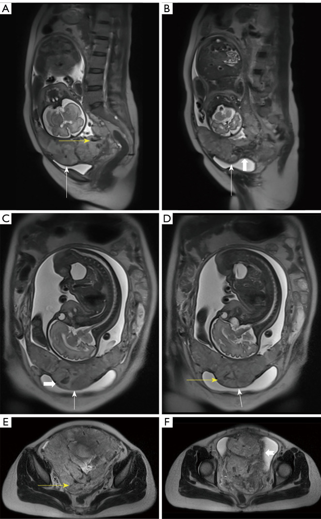
Figure 3.
A 37-year-old woman who had previously undergone two cesarean sections presented with complete placenta previa and concerns regarding placenta accreta spectrum disorder. The patient was at 30 weeks of gestation with placenta increta. The placenta extends to cover the anterior and the posterior walls of the uterus. (A,B) In sagittal, (C,D) coronal, and (E,F) axial half-Fourier acquisition single-shot turbo spin echo images, the presence of placental bulge is evident, characterized by widening of the lower uterine segment (white arrows). Additionally, T2-dark bands (white, thick arrows), abnormal intraplacental vascularity (yellow arrows), and myometrial interruption (arrowhead) were observed.

