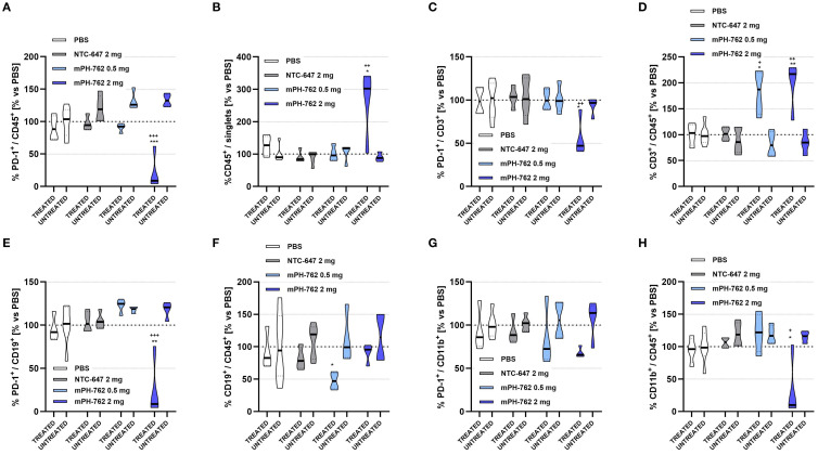
Figure 4.
Characterization of on-target immunomodulatory effects of intratumoral mPH-762 in the directly treated vs untreated distal tumor microenvironment isolated on in vivo study Day 14 by immunostaining/flow cytometry. Mice were implanted subcutaneously to bilateral flanks with 1e07 Hepa1-6 cells per flank, and treated IT (directly treated tumor only) with mPH-762, NTC or PBS on Days 1, 4, 7, 10 and 13. Violin plots are shown capturing data from each animal with medians indicated by treatment group for directly treated (DT) vs untreated distal tumors (UT). (A) Relative percentage (%) of PD-1+ / CD45+ tumor leukocytes in DT or UT each normalized to those of PBS treated animals (vs PBS). (B) Relative overall %CD45+ / singlet events in DT or UT vs PBS. (C) Relative %PD-1+ / CD3+ tumor T cells in DT or UT vs PBS. (D) Relative overall %CD3+ / CD45+ in DT or UT vs PBS. (E) Relative %PD-1+ / CD19+ tumor B cells in DT or UT vs PBS. (F) Relative overall %CD19+ / CD45+ in DT or UT vs PBS. (G) Relative %PD-1+ / CD11b+ tumor myeloid cells in DT or UT vs PBS. (H) Relative overall %CD11b+ / CD45+ in DT or UT vs PBS. Statistical significance of differences in mean AUC were assessed by one-way ANOVA and Tukey’s multiple comparisons post-hoc tests. ***p<0.001, **p<0.01, *p<0.05; + = vs NTC; * = vs PBS.

