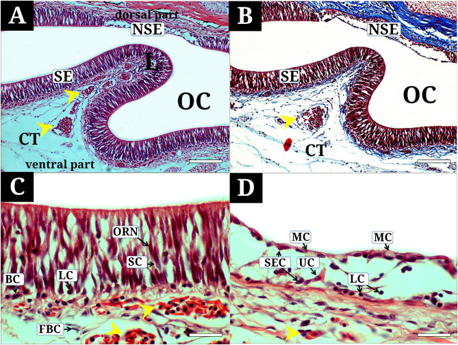
Fig. 3.
Histological characteristics of the olfactory epithelium of Synechogobius hasta, stained with hematoxylin and eosin (A, C, D), Masson’s trichrome (B). (A, B), the olfactory epithelium composed of sensory and non-sensory regions; (C) the sensory epithelium showing olfactory receptor neurons, supporting cells, basal cells, and lymphatic cells, and numerous blood capillaries and fibroblast cells in the connective tissue; (D) the non-sensory epithelium showing stratified epithelial cells, mucous cells, lymphatic cells, and unidentified cells. BC, basal cell; CT, connective tissue; FBC, fibroblast cell; L, lamella; LC, lymphatic cell; MC, mucous cell; NSE, non-sensory epithelium; OC, olfactory chamber; ORN, olfactory receptor neuron; SE, sensory epithelium; SEC, stratified epithelial cell; SC, supporting cells; unidentified cells, UC; yellow asterisk, blood capillary. The bars indicate 200 μm in A and B, 50 μm in C and D, respectively

