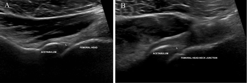
Fig. 7.
Long-axis US images during dynamic imaging for FAI with the transducer placed along the anterolateral aspect of the hip. (A) Image obtained at neutral and showing the anterolateral aspect of the normal, triangular-shaped acetabular labrum (L). (B) Representative, long-axis dynamic image obtained while flexing the hip to 90 degrees showing no evidence of FAI with no deformation of the labrum and no osseous/cam impingement

