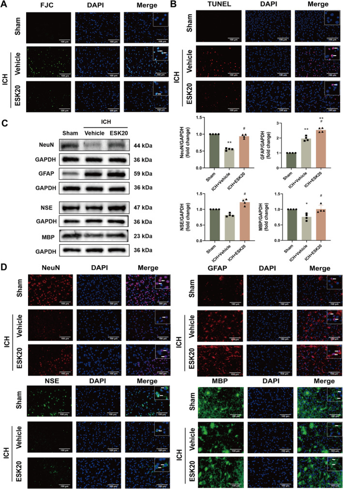
FIGURE 2.

ESK treatment reduces neuronal degeneration and death while increasing the protein expression of NeuN, GFAP, NSE, and MBP in mice subjected to ICH. (A) Representative images of Fluoro‐Jade C staining in the perihematomal region 72 h after ICH (n = 3). (B) Representative images of TUBEL staining in the perihematomal region at 72 h after ICH (n = 3). (C) Western blot analysis of NeuN, GFAP, NSE, and MBP protein expression, showing representative bands (n = 4), *p < 0.05, **p < 0.01 versus sham; # p < 0.05, ## p < 0.01 versus ICH + vehicle. (D) Immunofluorescence images of NeuN, GFAP, NSE, and MBP in the perihematomal region of mouse brain tissue after ICH (n = 3). Scale bar: 100 μm.
